BIOE Professor Publishes Global Consensus on Brillouin Microscopy in Nature Photonics
An innovative optical technique is one step closer to widespread clinical and research use, thanks to a global effort led in part by Fischell Institute Fellow and Professor in the Fischell Department of Bioengineering (BIOE) and at the University of Maryland and Co-Director of the the Edward and Jennifer St. John Center for Translational Engineering in Medicine (CTEM), Giuliano Scarcelli. This research, recently published in Nature Photonics, introduces the first internationally recognized standard for Brillouin Light Scattering (BLS) microscopy. BLS microscopy is a label-free, non-contact imaging technology that uses light to measure the mechanical properties of cells and tissues, such as stiffness and viscosity, at the nanoscale. These mechanical properties can indicate disease before traditional biological markers appear, making BLS a valuable tool in research and diagnostic settings. “Mechanical properties can tell us a lot about the state of a tissue or cell,” Scarcelli says. “With this consensus, we’re building the foundation that allows the field to grow with scientific rigor and clinical relevance.” Developed by a team of more than 40 researchers from institutions across the globe, the paper introduces a “Minimum Reporting Table” (MRT) to ensure consistency in how BLS experiments are conducted and shared. By laying out a standardized approach for the field, the work aims to make BLS microscopy more accessible, reliable, and comparable across labs worldwide, a critical step toward its adoption in biomedical research and clinical diagnostics.
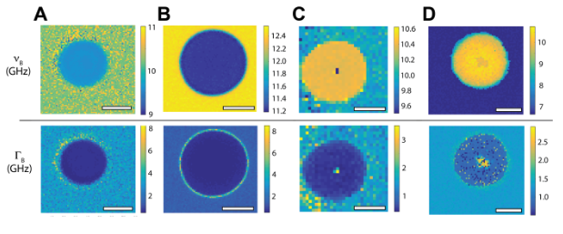
Figure 1: Spatial maps of Brillouin frequency shift (B) and linewidth (B) for a 10μm polystyrene bead with different Brillouin technology modalities. Scarcelli is Principal Investigator of the Optics Biotech Lab, where his team explores how light interacts with biological systems. His lab builds custom imaging tools that reveal hard-to-measure tissue characteristics like stiffness and mass, supporting clinical trials and translational science. Brillouin microscopy works by using light to detect natural acoustic vibrations in biological samples, revealing important mechanical characteristics such as stiffness and viscosity. These properties often play a key role in diseases like cancer, fibrosis, and eye disorders, making BLS a powerful tool for both basic science and translational medicine. With increasing interest from the medical community and technology developers, BLS is poised to become a mainstay in next-generation biomedical imaging. Scarcelli and his collaborators believe the new guidelines will accelerate both academic research and the commercialization of BLS-based diagnostic tools.
Related Articles: July 7, 2025 Prev Next |