Brain Freeze: Cryo-FIB-SEM Coming Soon to College Park
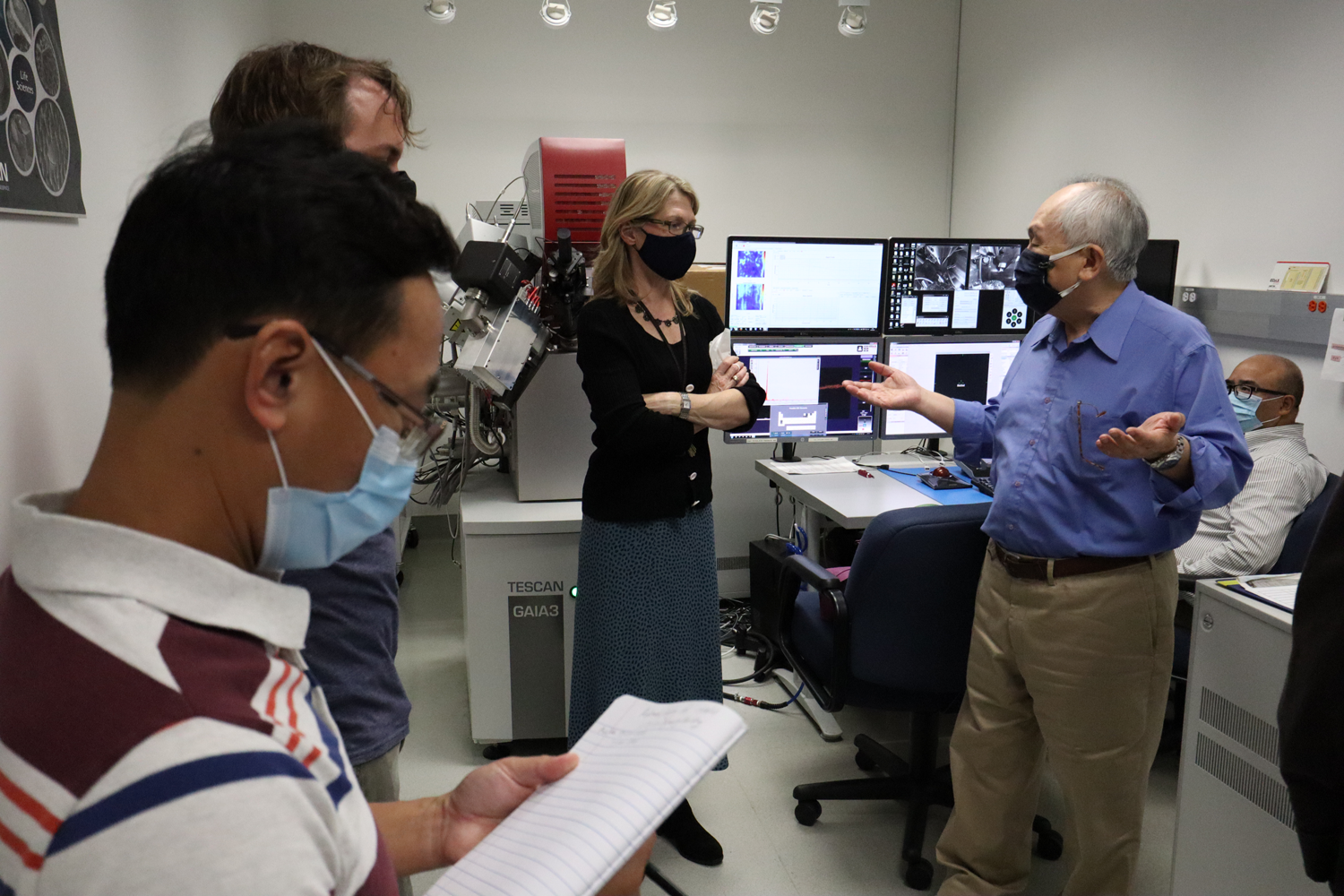
The Brain and Behavior Institute (BBI) is excited to announce that the University of Maryland’s focused ion beam scanning electron microscope (FIB-SEM)—an established tool in the physical sciences and materials engineering—will also soon support biological and neuroscience applications. This spring, the FIB-SEM located in the Advanced Imaging and Microscopy (AIM) lab at the University of Maryland’s Nanocenter will be outfitted with cryo- capability that allows for the three-dimensional reconstruction of biological ultrastructure in unfixed samples. FIB-SEM is a state-of-the-art block-face technology that repeatedly mills and scans a surface to allow for three-dimensional reconstruction of ultrastructure at high resolution. Under the direction of Wen-An Chiou, the AIM lab has employed FIB-SEM since 2015 for materials characterization and analysis at the nano- scale. And in 2022, neuroscientists and life scientists will be able to use cryo-FIB-SEM for high-fidelity imaging of biological processes in the absence of fixatives. “The expansion of the FIB-SEM to include cryo-preserved tissue from the nervous system will allow for 3D reconstruction of biological ultrastructure for the first time on campus,” said Elizabeth Quinlan, Professor of Biology, Clark Leadership Chair in Neuroscience and director of the BBI. “This powerful technique will support investigation of cytosolic and synaptic integrity, circuit connectivity and the ultrastructure of organelles or the extracellular environment.” For biological samples, the focused ion beam offers a closer shave than a traditional microtome, and this smaller z-step (with smallest milling depth of about 60-70nm) generates three-dimensional images with higher resolution and fewer defects. The tool combines the advantages of a focused ion beam with scanning electron microscopy, which possesses superior magnification for studying the structural details of synapses and other organelles in the nanometer range. Whereas the size of a typical mammalian synapse—about 1 micron—eludes the 1,000-2,000x magnification capabilities of conventional light microscopy, scanning electron microscopy is able to magnify structures between 100,000 and 300,000 times. “We are very excited to begin working with neuroscientists and other life scientists at UMD and from Baltimore,” said Chiou, “The cryo- capabilities are scheduled to be available as early as late spring, and FIB-SEM is currently available for fixed biological tissue right now.” For details on the current technology available at the AIM lab, please visit https://www.nanocenter.umd.edu/aimlab/. To set up a consulting appointment about how FIB-SEM and cryo-FIB-SEM can elevate your research, please contact Wen-An Chiou. ### Writer: Nathaniel Underland, underlan@umd.edu About the Brain and Behavior Institute: The mission of the BBI is to maximize existing strengths in neuroscience research, education and training at the University of Maryland and to elevate campus neuroscience through innovative, multidisciplinary approaches that expand our research portfolio, develop novel tools and approaches and advance the translation of basic science. A centralized community of neuroscientists, engineers, computer scientists, mathematicians, physical scientists, cognitive scientists and humanities scholars, the BBI looks to solve some of the most pressing problems related to nervous system function and disease.
Related Articles: January 31, 2022 Prev Next |


