UMD Research Group Develops New Technique for Detecting Elusive Protein Aggregates in Biopharmaceuticals
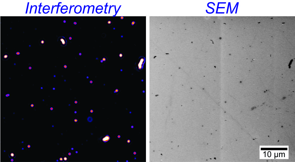
Monoclonal antibodies are therapeutic proteins used to treat a range of diseases that are unresponsive to traditional small molecule drugs, including various types of autoimmune diseases and cancer; melanoma, for example, is naturally resistant to drugs used in chemotherapy, which is part of why skin cancer is so dangerous. An inherent problem with these drugs is poor stability and shelf life. When exposed to excessive heat or shaking, for example, the monoclonal antibodies tend to clump together into microscopic protein aggregates – these aggregates pose a significant health risk to patients as they can cause unwanted immune responses, side effects, and reduced effectiveness of a potentially lifesaving drug. While there are a number of technologies for detecting protein aggregates in these drugs, they are currently limited to detecting larger protein aggregates with sizes of >1 micron (roughly 1/50th the size of a strand of hair). Enhanced methods are necessary to detect smaller aggregates, known as sub-micron protein aggregates, that are currently ignored in most analyses. In a study recently published in the Journal of Pharmaceutical Sciences, a research group in the Department of Chemical and Biomolecular Engineering (ChBE) at the University of Maryland developed a new optical microscopy based technique, called interferometric scattering microscopy (IFS), which can rapidly detect sub-micron protein aggregates. Nathan Wong, a ChBE undergraduate student and member of the Nanoscale Assembly and Electron Microscopy Lab, served as the paper’s first author. "By bouncing two light waves scattered off the particle and another reflective surface, we amplified the signal we received which let us see the particles with just a typical light microscope," Wong said. "Compared to other detection methods, IFS microscopy allows for much higher throughput - hopefully this will lead to a better understanding of how protein aggregates form." The main advantages of this approach are its simplicity, rapid measurement time (~10 minutes) and the detailed data it provides compared to existing techniques. Typical analytic methods require much more detailed analysis to detect sub-micron aggregates, while IFS only requires a simple sensor chip and benchtop optical microscope. “We established the resolution limit of IFS to be 100 nm protein aggregates and showed that the data it provides more accurately describes the population of aggregates compared with other existing analytical techniques,” said Taylor Woehl, ChBE Professor and PI on the study. “The really exciting aspect of this technique is that it uses light to observe sub-micron protein aggregates that would otherwise require much more complex analytical tools to detect.” The simplicity and rapid measurement times of this technique indicate it could be implemented in quality control labs at pharmaceutical companies to rapidly and robustly measure the amount of sub-micron protein aggregates, thus, contributing to improved safety of monoclonal antibody drugs. For additional information: Wong, N.A., Uchida, N.V., Dissanayake, T. U., Patel, M., Iqbal, M., T.J. Woehl (2019). “Detection and Sizing of Submicron Particles in Biologics with Interferometric Scattering Microscopy,” Journal of Pharmaceutical Sciences, DOI: 10.1016/j.xphs.2019.05.003.
Related Articles: July 25, 2019 Prev Next |